 Skip Navigation

Is it possible to take pictures of DNA with a cell phone camera?
June 7, 2024
- Related Topics:
- DNA basics,
- Molecular biology,
- Microscopy,
- Editor's choice
A teacher from San Francisco asks:
"Using my cell phone camera, I have been taking pictures of what I believe are DNA/genetic material. Is this possible? Images look like DNA strands to me!"
DNA is super small! It’s too small to see with your eyes, a camera, or even most microscopes.
It’s so tiny that you would need a very, very powerful special kind of microscope (like an electron microscope) to see a single piece of DNA.
But if you get a lot of DNA all clumped up together, it is possible to see with your eyes — or a cell phone camera!

A tiny instruction manual
Your DNA is essentially an instruction manual that makes you, you! It includes instructions for things like eye color, hair color, the ability to digest lactose in milk, and more.
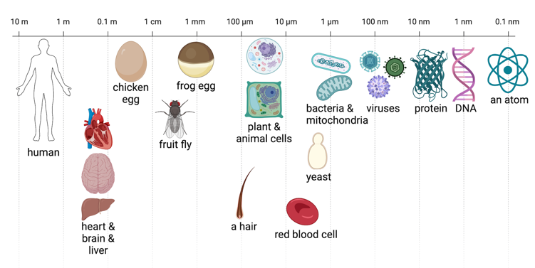
However, this instruction manual is teeny tiny. In fact, a single human hair is about 100,000 times wider than a piece of DNA, and a piece of DNA is about as wide as 10 atoms!
Picture this!
So how can we see DNA? Scientists have now developed special methods that allow us to take pictures of very tiny things, like DNA!
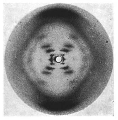
In 1952 Rosalind Franklin took the first picture of DNA. She did this by shooting X-rays at DNA and observing how the X-rays bounced off of the DNA. She then used the pattern that she saw in the X-rays bouncing off of the DNA to figure out the structure of DNA. This led to the discovery of the double helix shape that you probably imagine when you think of DNA.
While that image is very cool and very famous, it’s also hard for most of us to interpret, since it isn’t a direct picture of DNA. Instead, it’s a picture of how X-rays bounce off of DNA.
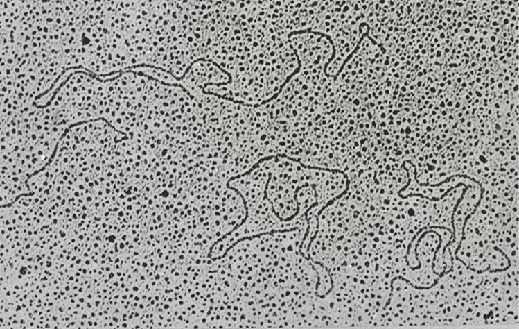
We can take direct pictures of DNA with electron microscopes. Those shoot electrons at it and measure the interactions between the electrons and the DNA. Even with these ultra-powerful microscopes, you usually can’t see the double helix itself. Instead, the DNA usually just looks like a line.
How can we take pictures of DNA with a cell phone?
While it is very cool that we can take pictures of DNA with a fancy microscope … most people don’t have one of those. So what kinds of pictures could we take with a cell phone?
The easiest option is to look at large amounts of DNA rather than individual pieces.
So how can we get large amounts of DNA? Let’s say you went to the farmers market last weekend and got a lot of strawberries, you just can’t eat them all and some are going to go to waste. Let’s extract their DNA instead of throwing them out!
First we can smash up the strawberries to start breaking open their cells that contain the DNA. Then we can add dish soap and water to finish breaking open the cells. We can then put this mushy mess into a coffee filter to just keep the juice.
Finally, we can force the DNA to “crash” out of the juice and form a big clump. Have you ever added sugar or salt to water and watched it disappear? To see our DNA we can do the opposite.
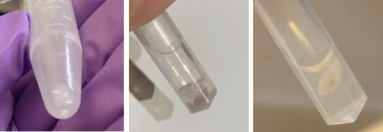
While the DNA starts out dissolved in water, if we add alcohol the DNA will un-dissolve and form a clump. The DNA will form a white stringy clump that you can see with your eyes … or a cell phone!
But remember that you can only see it because it is a ton of DNA, not just one piece.
Other cool stuff we can take pictures of with our cell phones!
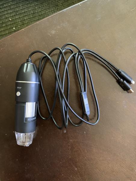
While writing this article I got inspired to see what other cool, tiny, stuff I could see with my cell phone! So, I got a cheap microscope attachment for my cell phone, and I set out to explore the environment around me to see what I could find.
Around the kitchen
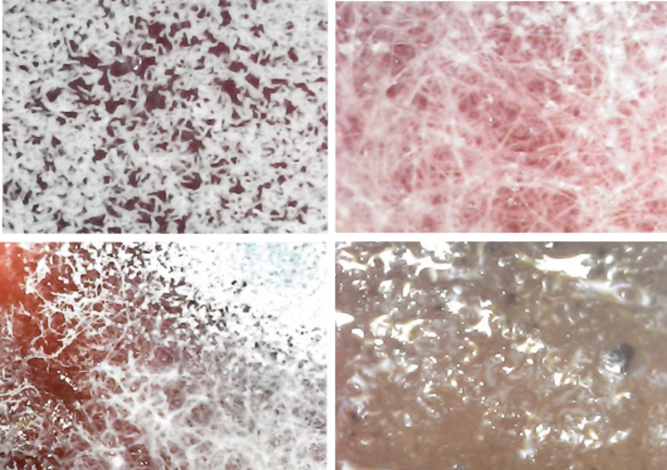
First, I found some old coffee sitting out on my friend’s counter that was beginning to grow fuzz and looked at the mold up close. There were a few different varieties of mold that looked different by eye, and they seemed to look really different under the microscope as well! Some had long stringy fibers while others had wider fibers. One didn’t have fibers at all and instead looked slimy and shiny.
Other fun stuff at home
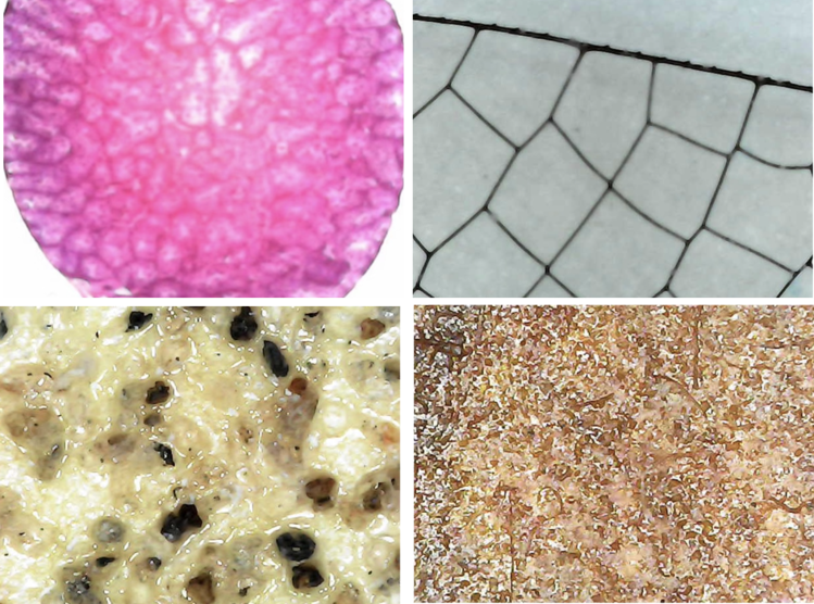
I also wanted to look at some common things that you might be able to find around the house. I had just completed a puzzle that was sitting on my coffee table, so I used my microscope to get a closer look. I was surprised to see that a puzzle piece that looked like a solid color by eye, was actually made of a bunch of different colored dots when I looked close up. I also looked at some carpet and found that I could see the individual fibers! Similar to what I found when I looked at a hand towel, but the pattern of the fibers was really different from the carpet. Finally, I found some bird feathers and found that different feathers looked different close up. Fluffier feathers had less defined lines up close, while smooth feathers had very well defined lines.

Pond water
Next I went dredging in a swampy area and grabbed some water full of pond scum. I found some cool critters living in the water! I found some worms and larvae and could watch them move around!

Plants
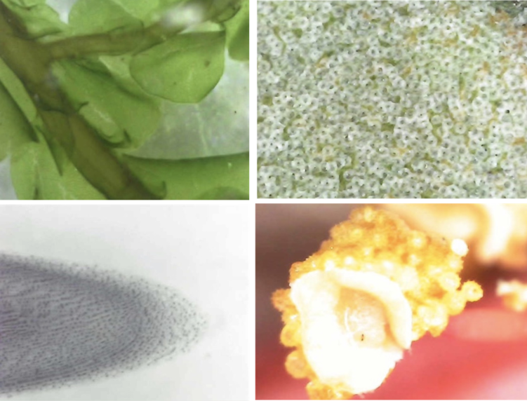
I also looked at some plants! I took some moss and looked at one little piece close up and could see each of the individual leaf-like structures. I also looked at the back of an olive leaf and found that there are little circular structures all over it. I could also see individual cells at the tip of an onion root! Finally, I picked a hibiscus flower from my garden and looked at the yellow pollen covered part from the center of the flower. When I zoomed in with my microscope I could see individual spikey, circular, pieces of pollen!
Around The Tech Interactive
Finally, I went exploring around the Tech with my microscope! I found so many cool things! I looked at a frog egg fixed on a slide. I could see the individual cells inside of the egg! I also saw a dragonfly wing and I could see the individual thread-like structures that make up the wing. I found a piece of sandpaper and when I zoomed in on it with my microscope I could see the individual pieces of sand in the sandpaper. Lastly, I looked at a dried SCOBY. A SCOBY is the microbial mat of bacteria and yeast that grows on top of kombucha while it is fermented and helps convert sugar to alcohol and gives kombucha its characteristic flavor. Up close the SCOBY had a lot of intricate structures that I wasn’t able to see by eye.
Read More:
- Can we see alleles with a microscope?
- King’s College: The story behind Photograph 51
- National Geographic: More on how microscopes work

Author: Danica Schmidtke
When this article was published in 2024, Danica was a PhD student in Ami Bhatt’s and Gavin Sherlock’s labs at Stanford University, interested in studying viruses that infect bacteria in the human gut and how they shape human health. Danica wrote this article while participating in the Stanford at the Tech program.